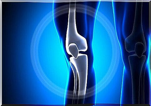
Knee Joint
Thanks to the knee joint, flexion and extension movements of the lower limbs can be performed. In addition, it is responsible for carrying out basic actions to walk, run and jump.
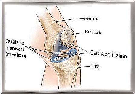
The knee joint is the largest in the human body and one of the most important, since it constitutes the point of union of three bones: the upper end of the tibia, the lower end of the femur and the patella.
Therefore, this joint serves as a communication element between the thigh and the leg. Do you want to know more about its anatomy? Next, we will tell you the most important functions of this part of the body and how it works. Do not miss it!
The knee joint, a biomechanical “wonder”
From what we mentioned earlier, there are those who affirm that the knee joint is a true “prodigy” of the biomechanics of the locomotor system. To understand this idea more, we must delve into its description and operation in parts, since the mechanics with which it operates is extremely complex.
The junction between the femur and the tibia forms the main body of this joint. Thanks to this, the knee supports the body weight; at the same time, the kneecap functions as a kind of pulley for the tendons.
This part of the body is endowed with great stability and this allows it to support the weight of the body. At the same time, it has a remarkable mobility to make walking and running possible, as well as to orient the foot according to the irregularities that a terrain may present.
Anatomy of the knee joint
There are several aspects that differentiate the knee joint from similar ones in the body. One of them is that it is a joint that, from a morphological point of view, is formed by the juxtaposition of two secondary joints :
- The patellofemoral: In it, the joint between the bones occurs by sliding. It protects the entire knee joint from the front and, at the same time, allows the quadriceps to be raised. In addition, it increases the pulling power of said muscle, since it places it at an angle that is conducive to it.
- The femorotibial: It is divided into two compartments or chambers, thanks to the articular meniscus. Such chambers are the proximal or superior and the distal or inferior. The first is responsible for the flexion and extension movements of the leg; meanwhile, the second allows rotational movements. It is also called the “meniscotibial joint.”
The femorotibial joint is bicolindal. This means that the two condyles of the femur, that is, the two rounded prominences that this bone has, roll freely on the tibia. More precisely, on an almost flat surface called the “tibial plateau”. There is no bone structure connecting these bones; they are tied together thanks to the ligaments.
Bony components of the joint
The bony components are the femur, tibia, and patella. According to an Arthritis-Helth post , these meet and move against each other at the knee joint. Next, we detail its main characteristics:
- Femur: bone almost cylindrical in shape, with a fairly bulging lower extremity. This gives you good support to transfer the weight to the upper part of the tibia. It has two condyles separated by a cleft called the “intercondylar fossa.”
- Tibia: articulates with the femur and supports the weight of the body, which is also transmitted to the foot. In the upper part, it has two cavities called glenoids, which house the condyles of the femur. Within these, the tibial spines are also found; there the cruciate ligaments are inserted. In the anterior part of the tibia, the patellar tendon is inserted.
- Patella: articulates with the femur in its posterior part. In the middle of both bones there is a cartilage called pre-patellar, which serves as a shock absorber between them.
Soft tissue components
Soft tissues are the tissues of the body that are not bones. According to information from the Cuban Health Network, the stability of the knee depends almost entirely on the supporting soft tissues, among which are the following:
- Joint capsule: it is a fibrous covering that surrounds the knee joint and forms a closed space. Inside, it joins the horns of the menisci; it is also connected to the tibia by the coronary ligaments.
- Synovial membrane: it is a thin layer that covers the joint capsule from the femur to the junction with the menisci.
- Bursa: Bursa are sacks filled with liquid. They act as if they were a cushion between the tendon and the bone. There are four: superficial, deep, prepatellar and tibiofemoral.
- Retinacles: are structures that allow the patella to connect with the menisci, tibia and femur. There are two: the medial and the lateral.
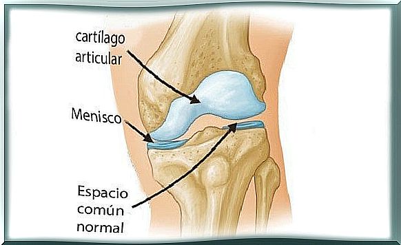
- Menisci: they are two fibrocartilages that are between the tibia and the femur and connect them. They are elastic and allow compression forces to be transmitted between the two bones.
- Ligaments: these are structures that give the knee stability and prevent extreme movements. There are intra-articular and extra-articular ligaments. Among the former are the anterior cruciate ligament and the posterior cruciate ligament; the latter include the internal lateral ligament and the external lateral ligament.
As you may have appreciated, the knee is a true work of art of nature. Among the thousands of biomechanisms that the human body possesses, the knee joint is, without a doubt, one of the most complex and functional. It is worth knowing how it works in order to take care of it in the right way.