The Diagnosis Of Pancreatic Cancer
Pancreatic cancer is a direct consequence of a defect in the division of the cells that make up this organ. They are divided into two types depending on the role they perform:
- Exocrine function: they produce enzymes that are released into the digestive tract and break down nutrients.
- Endocrine function: they produce various hormones that are released into the blood, for example insulin.
This is why pancreatic cancer can be of two types, exocrine or neuroendocrine. The diagnosis of pancreatic cancer differs between these two types, among other things, in the symptoms and signs.
Pancreatic Cancer Diagnosis by Signs and Symptoms
Depending on the lineage of the tumor, it will produce some symptoms or others.
Exocrine tumors
To understand them, a little explanation about the circulation of bilirubin is necessary. Bilirubin is a pigment that breaks down hemoglobin. Under normal conditions, it circulates through the bile ducts (from the liver to the intestine). Thus, it provides coloration to the stool.
The pancreas is located right next to the duodenum, which is where the bile ducts ultimately arrive. The growth of a tumor in this location causes obstruction. Thus, bilirubin stays in the liver, damaging it or passes to the skin or other organs. It is this that produces the symptoms listed below.
- To begin with, jaundice, which is the yellowing of the skin and mucous membranes.
- Acholia also appears, which are white stools, and coluria, which is brown-colored urine because bilirubin is excreted by the kidneys.
- It can cause itchy skin.

The above symptoms generally do not occur in neuroendocrines because they grow less. The growth and invasion of structures also causes stomach and back pain. Nausea and vomiting are also experienced. Nonspecific symptoms of cancer are weight loss and poor appetite.
Neuroendocrine tumors
Depending on which hormone is produced by the cell from which the tumor develops, some symptoms or others will be experienced.
Imaging tests
The following tests are done to diagnose pancreatic cancer:
Computed tomography (CT or CAT)
By means of this test, generally cross-sectional images are obtained using X-rays. There is a special type of CT to observe the pancreas, which is the pancreatic protocol CT. During it, an intravenous contrast is introduced that reveals the areas that are most interesting to see.
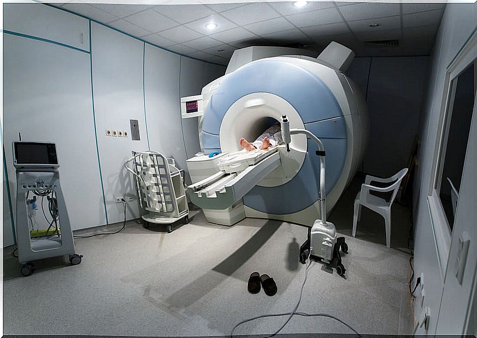
Magnetic resonance imaging (MRI)
In this case, images are obtained by subjecting the body to radio waves and magnetic fields. There is also a special MRI, which is MRI cholangiopancreatography, by which bile and pancreatic ducts are observed. With it we could show a possible obstruction.
Ultrasound
It is a technique that makes it possible to observe each structure inside the body by detecting the density of each one. This may be the first test to be done because it is quick and easy and does not involve exposing the person to radiation.
In the case of the pancreas, it may be less useful than previous tests as it offers poorer definition. However, an endoscopic ultrasound can be done, placing the ultrasound at the end of an endoscope (a flexible tube that is inserted into the intestine).
Taking the image from this point, very close to the pancreas, can greatly increase the definition of the image. It is also possible to associate a biopsy with an endoscopic ultrasound, accessing the tissue from this adjacent location.
Endoscopic retrograde cholangiopancreatography (ERCP)
In the course of this technique, the endoscope is introduced to the common bile duct, which runs from the bile and pancreatic ducts to the beginning of the intestine.
From here, the bile and pancreatic ducts are accessed and a contrast is introduced that makes it possible to see them better. Through this test we can see if any of these ducts is obstructed by a possible pancreatic tumor.
Somatostatin receptor scintigraphy or “octreoscan”
In this case, a hormone analog (octreotide) attached to a radioactive substance is injected. This set adheres to the tumor and is highlighted when the image is taken, but only in the case of neuroendocrines.
Positron emission tomography (PET)
It uses a mechanism analogous to the previous one, but this time glucose bound to a radioactive substance is injected. The tumor, having a lot of cellular activity, consumes a lot of glucose, and this is reflected in the image. Today, a combined image between PET and CT can be taken, greatly increasing the definition.
Analytical tests
The following analytical tests are also commonly performed:
Exocrine tumor
Liver function laboratory values may be altered. The levels of some tumor markers can also be found to be elevated, such as:
- Carcinoembryonic antigen (CEA)
- Ca 19.9
Neuroendocrine tumors
In this case, the levels of certain hormones such as insulin, gastrin, glucagon, somatostatin, pancreatic polypeptide and vasoactive intestinal peptide (VIP) may be altered.
Biopsy for the diagnosis of pancreatic cancer
A sample of the pancreas can also be taken and later analyzed in the medical department of Pathology and thus determine if it is tumor or not. This can be done percutaneously (through the skin), endoscopically, as discussed earlier, or surgically.